A closer look at microscopes
Logo

Nobel Prize monthly, April 2026 

Human stem cell

Human stem cell embedded in a 3D matrix, Cryo SEM. Credit: Sílvia A Ferreira, Cristina Lopo and Eileen Gentleman, KCL. Source: Wellcome Collection. Attribution 4.0 International (CC BY 4.0)

Revealing secrets on a tiny scale 

The microscope’s ability to be an extension of the human eye has enabled scientists to make strides in biology and medicine since its invention in the 16th century. Microscopes have had a huge impact on our understanding of everything from the composition of materials to the building blocks of life. Learn more about the work of some of the Nobel Prize laureates that have allowed microscopes to evolve. 

Frits Zernike

Frits Zernike with a phase contrast microscope. Courtesy of University of Groningen

Revolutionised medical research 

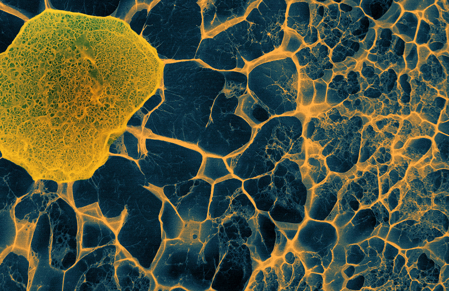
When light rays pass through transparent materials, such as biological specimens, there is a change in the phase of the light waves – the position of the wave crests in relation to one another – compared to an unimpeded light ray. Our eyes can’t detect this but these phase changes contain important information that can be used to visualise the material they have passed through.


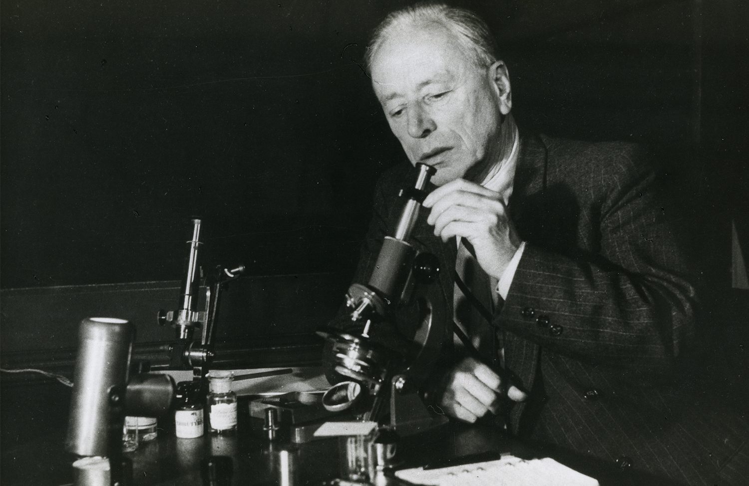
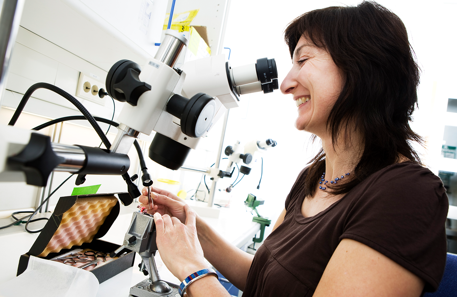
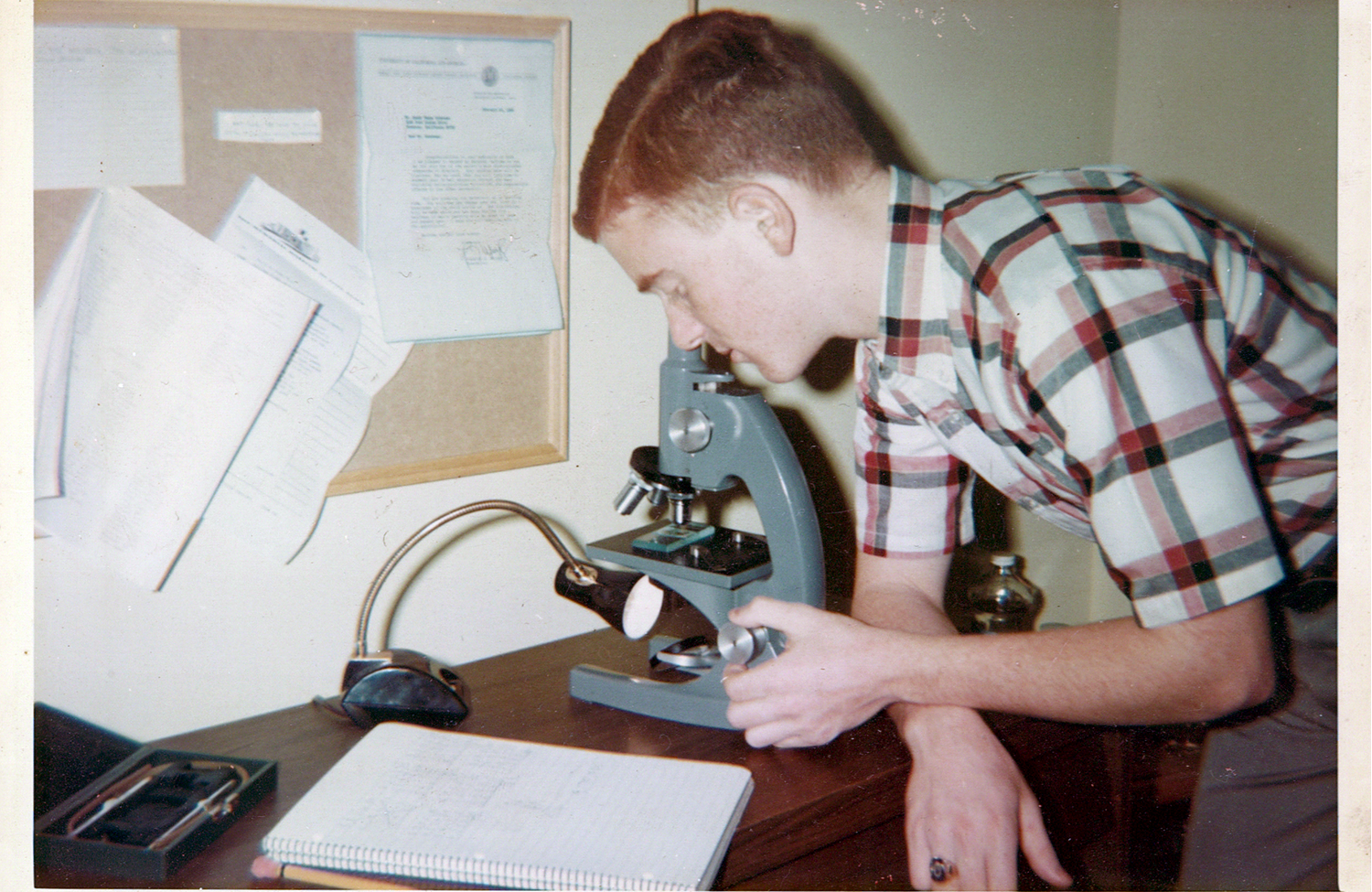
In the 1930s, Frits Zernike used this information to build a new scientific tool – the phase contrast microscope. His new microscope enhanced the contrast of unstained, transparent specimens to reveal their inner workings in richer detail. Phase contrast microscopes have enabled strides in biological and medical research to be made, and they allow oils, drugs and textiles to be scrutinised. Read more

May-Britt Moser

May-Britt Moser. Photo: Geir Mogen, NTNU (CC BY-NC 2.0). Attribution-NonCommercial 2.0 Generic (CC BY-NC 2.0). Photo taken from: https://flic.kr/p/avEtP2

“You see thousands of cells glowing up like stars”

Microscopes continue to enable revolutionary discoveries. May-Britt Moser was awarded the Nobel Prize in Physiology or Medicine 2014 for her discovery of so-called grid cells in the brain, that make it possible to determine position and to navigate. In this conversation, she speaks about her research and how she decided to share her knowledge with the science community. Watch here

De-mystifying mixtures

In chemistry, very small particles that are finely dispersed in another substance are called colloids. In 1902, Richard Zsigmondy introduced his Nobel Prize-awarded idea that led to the ultramicroscope, which made it possible to observe small colloidal particles not visible in a conventional microscope. The method can be compared to how dust particles suspended in the air sometimes become visible if we happen to be standing to the side of a window on a sunny day.

Light shines through a window

Guilherme Moreira, CC0, via Wikimedia Commons

More than a century later, applications of colloidal particles are abundant, with the most spectacular being the use of LNPs (lipid nanoparticles) in the COVID vaccine to transport mRNA to cells. Read more

“If the molecules are different, you can tell them apart”

In 2014, Stefan Hell was awarded the Nobel Prize in Chemistry for a method that improved the microscope resolution by orders of magnitude relative to the Zsigmondy method (see above).

Stefan Hell

Stefan Hell in the laboratory. Photo © Max Planck Institute for Biophysical Chemistry

In this video, Stefan Hell explains the basis of his discovery, the super-resolution fluorescence microscope, which allowed scientists to see sharper pictures of living cells and tissue than ever before. Watch here

Randy Schekman

Randy Schekman working on his Annual Science Fair project during his senior year of high school in the spring of 1966. Photo: Courtesy of Randy Schekman.

“I wanted to run away from home”

Aged 12, Randy Schekman was determined to save money from odd jobs to buy his first professional microscope. But he couldn’t reach his goal as his mother kept borrowing money from his piggy bank. “One Saturday I became so upset that, after mowing a neighbour’s lawn, I bicycled to the police station and announced to the desk officer that I wanted to run away from home because my mother took my money and I couldn’t use it to buy a microscope,” he said.


The desperate measures paid off: his parents picked him up from the police station and bought the microscope on their way home. Read Schekman’s biography

A new era of biochemistry

Thanks to the work of Richard Henderson, Joachim Frank and Jacques Dubochet, researchers can now freeze biomolecules mid-movement and portray them at atomic resolution. Cryo-electron microscopy builds on Henderson and Frank’s research using electron microscopes to visualise biological samples and Dubochet’s method of cooling the water around molecules so rapidly they can be imaged in their ‘natural’ form.

Illustration of the Zika virus

Visualisation of the Zika virus, enabled by cryo-electron miroscopy. © Johan Jarnestad/The Royal Swedish Academy of Sciences

In medical research, the technique can, for example, observe the 3D outer shell structures of viruses and determine the structure and interactions of proteins that play a significant role in cancer. Read more

Monthly quiz

A fascinating structure

On this day, 8 April, in 1982, Dan Shechtman looked at a picture produced by his electron microscope and couldn’t believe his eyes. He saw a pattern of concentric circles, each made of ten bright dots at the same distance from each other. The picture told Shechtman that the material he was studying had a crystal structure that had never been seen before, one that did not repeat itself, and was considered to be impossible. What is the name of Shechtman’s Nobel Prize-awarded discovery?

Kip Thorne

Photo: Alexander Mahmoud

This email was sent to you as a subscriber of Nobel Prize monthly

We maintain a strict privacy policy for the newsletter. When you sign up, you can be assured that your email address will not be sold or given to any third party.